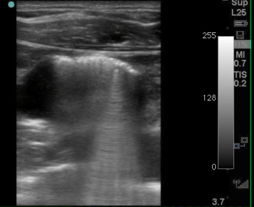
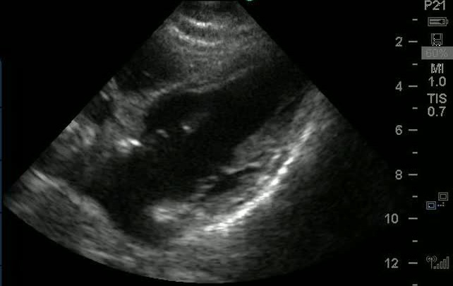
Improving your echocardiograms with contrast agents

2016-10-13T18:16:29-05:00October 14th, 2014|Ultrasound|

Echocardiograms can often be limited by poor sonographic windows, but using an echo contrast agent can improve visualization of the left ventricle and detect right-to-left intracardiac shunts. Introduction to the various agents, their principles and use.

Air visualized within IJ after penetrating trauma

2016-10-13T18:16:37-05:00April 25th, 2013|Trauma, Ultrasound|

Take a look at this really remarkable ultrasound image, not so much for it immediate clinical utility, but for its rarity. A patient presented with numerous self-inflicted cuts all over the body including bilateral extremities, torso, and neck. Arrived profusely bleeding, and clear

Ultrasound Guided Ankle Arthrocentesis

2016-10-13T18:16:39-05:00March 27th, 2013|Orthopedics, Ultrasound|

My preferred method is the anterior-medial approach. Place the patient in the supine position with knee flexed, and foot plantar flexed and resting flat on the bed. An high-frequency linear probe is most useful for identifying the detailed structures. Using a combination of palpati

Algorithm for Evaluation of Non-apparent Globe Injuries

2016-10-13T18:16:39-05:00March 11th, 2013|Trauma, Ultrasound|

Globe rupture is more often occult on presentation. The most frequent sites of rupture are not easily visualized, and more superficial injuries may block examination of the posterior segment. Very small sharp foreign bodies can enter the eye through small wounds that are difficult to visualize. Our institution has developed the following protocol for evaluation of clinically non-apparent ruptured globe injuries.

Ultrasound demonstrating Incompetent Cervix

2016-10-13T18:16:41-05:00February 21st, 2013|Ultrasound|

This ultrasound is from a young pregnant woman presenting with lower abdominal pain without vaginal bleeding. History of two previous unexplained spontaneous abortions and a known large fibroid. The physical exam was unremarkable but bedside transabdominal ultrasound was performed