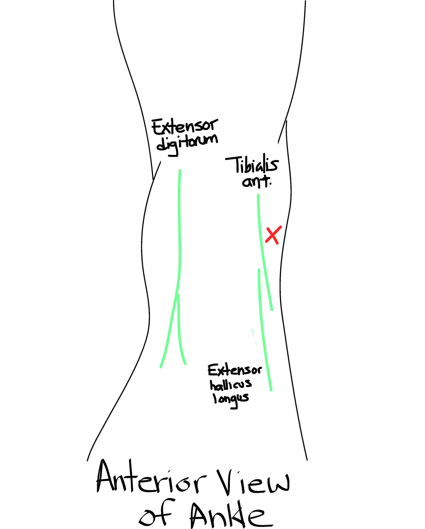
My preferred method is the anterior-medial approach. Place the patient in the supine position with knee flexed, and foot plantar flexed and resting flat on the bed. An high-frequency linear probe is most useful for identifying the detailed structures. Using a combination of palpation and ultrasound, identify the anatomical landmarks including the Extensor digitorum, tibialis anterior, Extensor hallicus longus, and the doralis pedis artery (which is deep and lateral to to the extensor hallicus longus).


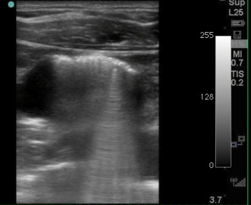
The effusion appears as an hypoechoic structure just distal to the tibia in the tibiotalar recess. The probe should be medial to the tibialis anterior tendon. The procedure should be performed in sterile conditions.
The fluid should be sent for cell count and differential, culture, and crystal analysis. An interesting method of distinguishing septic arthritis, from underlying gouty arthritis may be the measurement of lactate (Synovial Lactic Acid and Septic Arthritis. JAMA. 2007;298(1):37).


Contribute your thoughts.