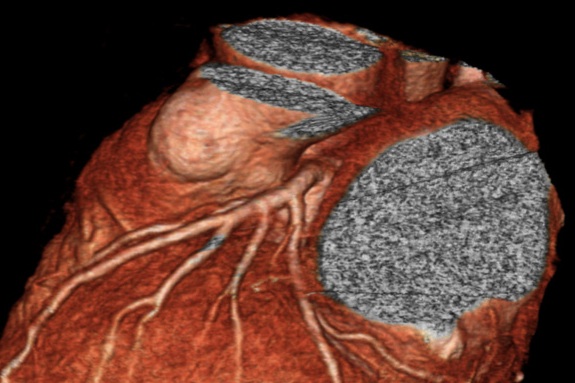
CT evaluation of the coronary anatomy (CT coronary angiogram) has developed rapidly as scanner technology and software have made dramatic leaps forward in capability. The introduction of multidetector scanners allows for capturing a temporal sequencing needed to image a beating heart, and the resolution allows for the imaging on the scale of the epicardial coronary arteries. The specific scanning protocols have also advanced reducing the patient radiation exposure. All of these factors now allow for the CT coronary angiography to be a powerful tool in the evaluation of of a patient with suspected acute coronary syndrome. The following is a protocol developed at our institution for the emergency department to use in preparing the patient to for a CT coronary angiogram.
Indications to obtain CT coronary angiogram
- Evaluation of chest pain syndrome or suspected cardiac ischemia.
- Must be of sufficient risk that, without the test, you would admit to observation.
- Should be for patients age < 55 (men) <65 (women), or < 45/55 if they have 3 CAD risk factors, or known CAD. Age and risk factors are general guidelines, and exceptions can be made. Exception is also appropriate for very atypical pain or patients with a previous negative or indeterminate stress test. You may use your sound clinical judgment.
- An initial negative troponin is recommended but not necessary.
How much radiotaiton exposure is there?
- 3-5 milliseverts of radiation on the 256 slice scanner.
- Only 10 minutes for reconstruction.
- Only 85 cc contrast (formerly 100cc); working toward 70 cc contrast.
Screening Questions (all must be no)
CAD risk Stratification
- High suspicion of acute coronary syndrome based on history/exam.
- Significant ischemic ECG findings.
- One troponin elevation above the 99th percentile on your assay (consider a second troponin after CT, before discharge).
- In the case of prolonged constant pain > 12 hours, one negative troponin is sufficient.
- Prior invasive cardiac catheterization.
- CTA within the last year.
- Patient has three or more of the following cardiac risk factors: diabetes, hypertension, family history of premature atherosclerosis, dyslipidemia, tobacco use.
Contraindications to the test
- Presence of atrial fibrillation or frequent PVCs, PACs.
- Is unable to hold breath for 10 seconds or follow instructions.
- Known allergy to intravenous contrast.
- Weight greater than 250 pounds.
What laboratory tests are required before the CT coronary angiogram?
- If the creatinine is ≥ 1.5, test may not be ordered without cardiology/radiology clearance.
- Bicarb protocol: If GFR >60, no bicarb. If GFR 45-60, bicarb only if risk factors present, GFR 30-45, bicarb regardless of risk factors, GFR <30, should not be giving contrast.
- Risk factors: DM, HTN, prior kidney disease, age >70 years.
How to prepare your patient for the test
- Heart rate ideally < 65 and regular.
- Patient should be NPO four hours prior to the examination except medications.
- Intravenous Access must be: 18 gauge right antecubital fossa (left is ok if right unavailable) IV.
- Beta-blockade (metoprolol):
- Contraindications: SBP<100, active wheezing, known CHF
- Oral
- Pulse 65-75: 50 mg po, scan 45 minutes later
- Pulse > 75: 100 mg po, recheck in 45 minutes
- Intravenous
- If HR still >75, administer up to 30 mg IV metoprolol in 10 mg increments
- After maximum metoprolol given, if HR <75, send patient for scan. If >75, cannot scan
- Contraindication to metoprolol: give oral rapid absorbing diltiazem 60-90 mg.
Disposition based on Results
- No plaque: Discharge
- Calcium > 400 — No contrast, admit
- Any stenosis > 50% — Admit with Dx of ACS
- Any stenosis 0-50% — Check 2nd troponin, Aspirin, Cards clinic ASAP
- Soft plaque — Check 2nd troponin, Aspirin, Cards clinic ASAP
- Signif calcium (100-400) — Aspirin, Cardiology clinic ASAP vs. admit
This protocol was developed at Hennepin County Medical Center by Drs. Stephen Smith, Gopal Punjabi, Bradley Bart, and Woubeshet Ayenew. While this protocol is in use at our institution and can serve as a guide, it should be individualized at each center with consultation between Cardiology, Radiology, and the Emergency Department.
Contribute your thoughts.