Critical aortic stenosis limits cardiac output and can be life-threatening in patients who develop significant anemia. Oxygen delivery to tissues is inadequate. This can be seen in an elderly patient with calcific aortic valves who suffers an acute gastrointestinal bleed such as from ulcerations, diverticular bleed, or bleeding from a vascular malformation.
Unfortunately, the aortic stenosis itself may be the mechanism leading to the anemia.
The anemia may develop by two mechanisms:
- Intravascular hemolysis-dependent anemia occurs by turbulence and shear stress across a mechanical or narrowed calcified valve.
- Acquired von Willebrand syndrome. Shear stress in this case causes destruction of vWf multimers, and low-grade chronic hypoxia causes smooth muscle relaxation and reflex sympathetic vasodilation leading to ectasia of blood vessel walls. When combined, this can cause slow oozing hemorrhage which can lead to significant decreases in hematocrit.
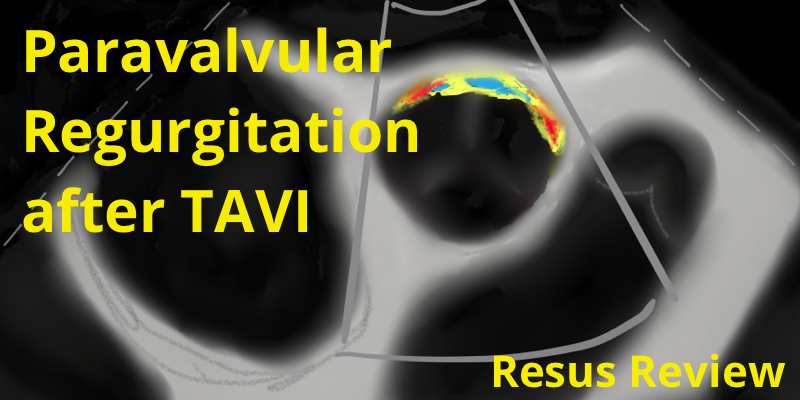
Heyde’s syndrome is the triad of significant aortic valve stenosis, acquired coagulopathy, and subcutaneous, mucosal, or gastrointestinal angiodysplasia dependent anemia.
The bleeding that occurs with the effective platelet dysfunction is mucosal. Platelets function well enough that petechiae and purpura do not occur.
No simple, single laboratory test is available to screen for vWD, and the diagnosis is made using several specific laboratory tests. Screening tests typically include prothrombin time (PT), activated partial thromboplastin time (aPTT), F VIII coagulant activity, ristocetin cofactor (RCoF) activity, and concentration of vWF antigen (vWF:Ag). It is important tha vWF levels are compared against ABO specific normal ranges for your laboratory.
Contribute your thoughts.