Fiberoptic oral intubation with flexible fiberoptic endoscope is a useful technique for establishing an airway in patients with abnormal upper airway anatomy, cervical immobilization, or awake intubations with preservation of the patient’s respiratory drive to when it is desirable to maintain spontaneously breathing during the intubation. Unlike endoscope or blind nasotracheal intubations, an oral approach can be more challenging because the scope must make a sharp turn in the posterior oropharynx before entering the hypopharynx. During nasotracheal intubation, the scope can make the turn in the nasopharynx and take a straight path towards the trachea.
It is key that patient is adequately anesthetized. Sedation with medications that allow spontaneous breathing and allow the patient to protected the airway are useful, avoiding deep sedation and paralytics as used in RSI. These can include ketamine, droperidol, dexmedetomidine, etc.
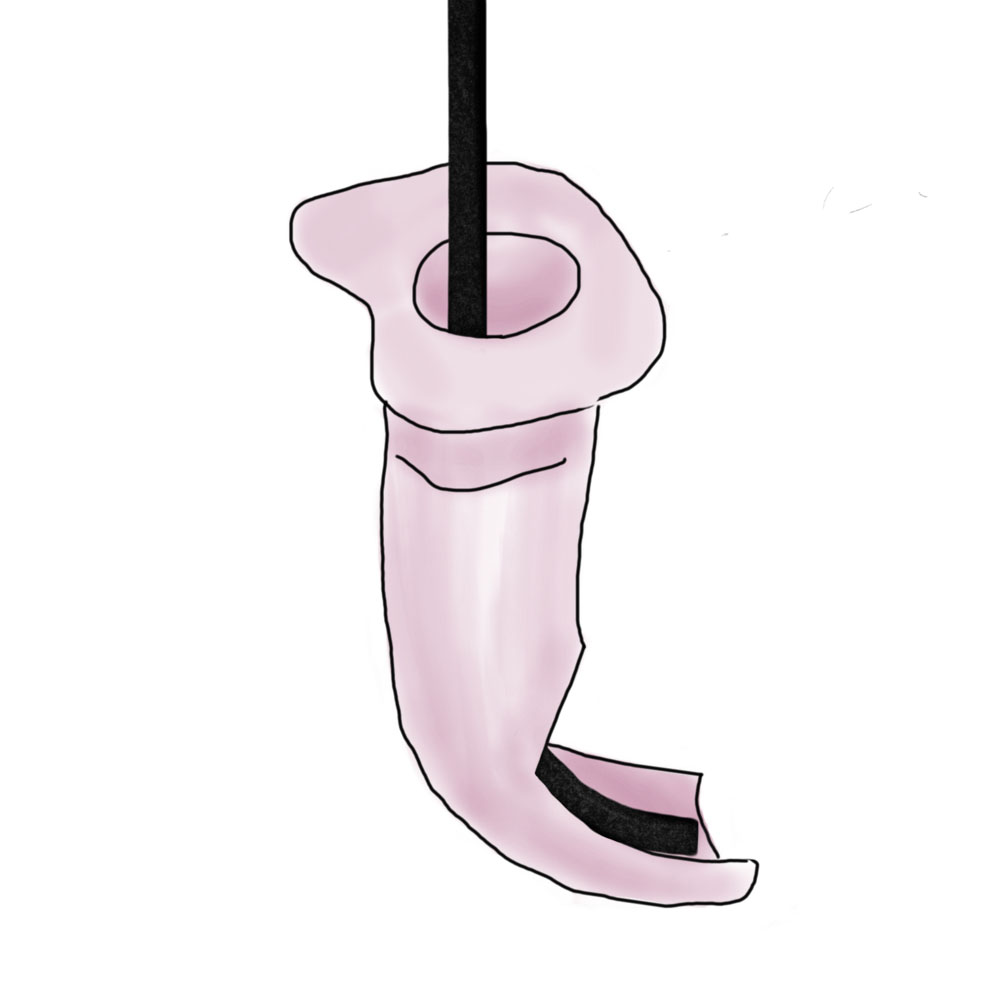
Oral Airways for Intubation
Since the patient is generally in an upright position, the tongue, mandible soft tissues can fall backwards obstructing the airway. This can make passage of the fiberoptic scope difficult. This can be improved with a jaw thrust, but is facilitated greatly by the use of a specialized intubating oral airway.
There are three available oral guides (bite protectors) including the Williams airway (shown below), Berman airway, and Ovassapian airway. They are all specifically designed for oral fiberoptic intubation to guide the fiberoptic scope and endotracheal tube around the oropharynx and keep the tongue and hypopharyngeal tissue from obstructing the trachea. The airways also protect the scope from being damaged if the patient bites down. The Williams airway comes in a 9cm “female” size and 10cm “male” size. Well lubricated, a 6.5 ETT can pass through either. Remember, after you have intubated, you must remove the 15mm adaptor to be able to remove it.
Equipment
- Sedative medication
- Fiberoptic scope (eg bronchoscope)
- Lidocaine 4% cream and tongue blade
- Lidocaine nebulization
- Appropriately sized endotracheal tube
- Williams airway
- 10 mL syringe
- Securing device for endotracheal tube
- Capnography monitor / Colorimetric device
Once you have gathered all of the equipment and the patient has been adequately preoxygenated, you can begin the actual intubation steps.
Procedure Steps
- Remove 15mm adaptor form endotracheal tube and on fiberoptic scope.
- Place patient in comfortable sitting or near sitting position.
- Lidocaine nebulization.
- Lidocaine 4% cream placed on the back of the patient’s tongue. Have the patient gargle the cream as it runs down the back of their mouth.
- Ketamine dissociative medication.
- Insert the Williams airway.
- Advance the scope to the end of the Williams airway.
- Assistant can make minor adjustments to the position of the Williams airway that scope it is directed at the vocal cords.
- Inject lidocaine 4% liquid on cords.
- Advance the scope through the vocal cords and into the trachea.
- Advance the endotracheal tube over bronchoscope through the vocal cords.
- Remove the fiberoptic scope and the Williams Airway.
- Replace the 15 mm adaptor
- Inflate the endotracheal tube balloon and confirm placement.
- Provide patient extra sedation if required.
Contribute your thoughts.