Defining the coronary artery anatomy is a critical step in any evaluation of ischemic heart disease and developing a treatment plan for your patient.
The location of atherosclerotic lesions can be suggested by provocative stress testing (exercise or pharmacologic stress, and multiple evaluation modalities including ECG, echocardiography, and nuclear medicine). However, definitive identification can only be accomplished by either left heart catheterization or CT coronary angiogram. While there are benefits to both tests, catheterization defines lesions geometrically and functionally, and during the same procedure allow intervention (PCI) if indicated.
Major Coronary Arteries
The coronary anatomy is complex and varies significantly between patients (eg. example of different origin). The normal major epicardial arteries are shown in the figure below.
The left coronary cusp gives rise to the left main coronary artery which branches to the left anterior descending (LAD) and left circumflex artery (LCx, Circ). The branches from the LAD are labelled in sequence diagonal 1, diagonal 2, etc. Similarly, branches from the LCx are labelled obtuse marginal and numbered in order. The LCx travels in the left AV groove, while the LAD is a large anterior artery in the interventricular groove.
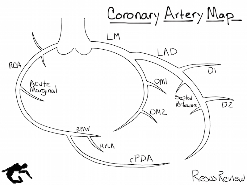
The right coronary artery (RCA, Right circumflex) is the only major artery that originates from the right coronary cusp. It travels in the right AV groove to the posterior inferior part of the heart. In 80% of patients, it then turns towards the apex in the interventricular groove as the posterior descending artery (PDA).
Diagramming Coronary Anatomy
The coronary arteries form a three-dimension structure, and there are various ways to represent them on a 2-dimensional diagram. Like maps, the various diagrams emphasize different aspects. Some are more conceptual, others focus on branching, while still others attempt to preserve a spatial representation.
I developed the diagram below specifically for markup purposes. It sacrifices geometry for ease of interpretation and diagramming. Wide spacing and large arteries allow you to easily annotate lesions and surgical grafts.

Download Coronary Artery Diagram pdf.
Marking Up Coronary Artery Diagram
Understanding your patient’s coronary anatomy can be difficult to visualize, especially when there are multiple lesions, interventions, and possibly coronary artery bypass surgery (example of multiple lesions and by pass grafting).
Using the coronary artery diagram, you can track lesions, stenting, grafts, size size/variation of native arteries. The example shown below, shows two locations with in-stent restenosis, three new lesions, a chronic total occlusion, collaterals between the LAD and LCx, and a very large D1 vessel.
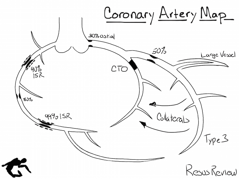
I find this useful while getting a history of the patient, interpreting cardiac catheterization reports, and reading surgical records. This documentation then helps with preop planning and facilitating communication between teammates.
Cardiac catheterization labs are moving towards structured reported (rather than solely textual), which can then be part of the permanent medical record.
[…] From ResusReview […]