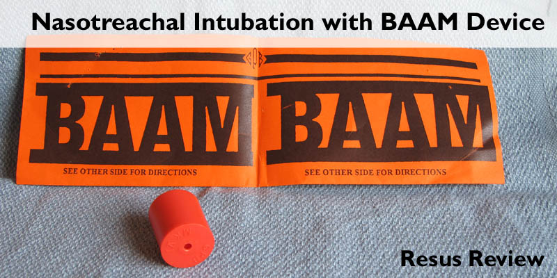
Ultrasound Guided Ankle Arthrocentesis

2016-10-13T18:16:39-05:00March 27th, 2013|Orthopedics, Ultrasound|

My preferred method is the anterior-medial approach. Place the patient in the supine position with knee flexed, and foot plantar flexed and resting flat on the bed. An high-frequency linear probe is most useful for identifying the detailed structures. Using a combination of palpati

Basics of Soft Tissue Neck Injury

2016-10-13T18:16:39-05:00March 21st, 2013|Trauma|

Zones of the Neck Defined by mandible, cricoid cartilage, and sternal notch. Most injuries sustained in Zone II, especially carotid injuries Zone I mortality high due to intrathoracic injuries. Posterior triangle with few vital structures. Important exception is subclavian at risk

Fullminant Tuberculosis

2016-10-13T18:16:39-05:00March 17th, 2013|Pulmonary|

Tuberculosis is an endemic problem through most of the world. However, when it presents as active pulmonary disease in the United States, especially in nonimmigrrant, it is usually early in the disease process. This patient a native resident, had delayed seeking care for months. The c

Algorithm for Evaluation of Non-apparent Globe Injuries

2016-10-13T18:16:39-05:00March 11th, 2013|Trauma, Ultrasound|

Globe rupture is more often occult on presentation. The most frequent sites of rupture are not easily visualized, and more superficial injuries may block examination of the posterior segment. Very small sharp foreign bodies can enter the eye through small wounds that are difficult to visualize. Our institution has developed the following protocol for evaluation of clinically non-apparent ruptured globe injuries.

Bennett’s Fracture

2016-10-13T18:16:40-05:00March 10th, 2013|Orthopedics|

Fracture of the base of the first metacarpal bone which extends into the carpometacarpal joint. This intra-articular fracture is the most common type of fracture of the thumb. Caused by an axial force directed against the partially flexed metacarpal, such as during a fight or fall

Lead Placement for Posterior ECG

2016-10-13T18:16:41-05:00March 10th, 2013|Cardiology, ECG|

The leads V4-V6 are removed and substituted for V7-V9 as shown below. On most EKg machines, the labels areno automatically changed so it is important to cross out the labels for V4-V6 and write in V7-V9. It is also helpful for future clinicians, if you note in your read that it is a p